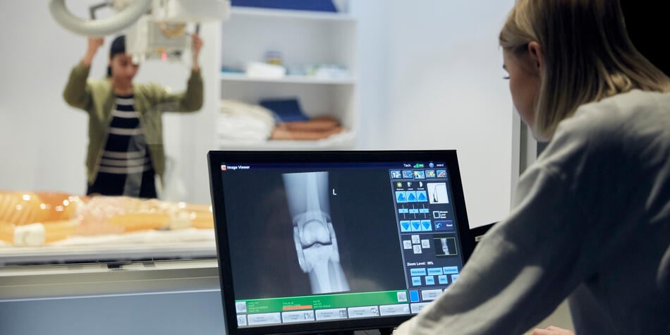
Do you get a buzz working with the latest technology? Thrive working in cross-disciplinary teams and keen to make a real difference in a healthcare setting? A career in medical radiography might be perfect for you.
‘X-ray machines and other medical imaging technology is upgraded every three or four years, so if you’re someone who likes technology, science and working with people, it’s an ideal choice,’ says Associate Professor Giovanni Mandarano from Deakin University’s School of Medicine.
What is a radiographer?
Radiographers use different types of radiation to produce high-quality medical images – like x-rays, CT scans and MRIs – to help specialists and other health practitioners describe, diagnose, monitor and treat injury or illness.
‘If you’ve ever had a broken bone, you’ve probably had an x-ray,’ says Assoc. Prof. Mandarano. ‘A CT scanner uses x-rays and digital computer technology to create detailed images of the body, while an MRI scan uses magnetic fields and radio frequency waves to take pictures inside the body. It’s excellent for looking at the details in the brain and spinal cord.’
CT and MRI scans can reveal tumours, trauma and spinal injury, as well as assess the structure and shape of body parts. With the aid of medical imaging doctors can make informed diagnoses and treatment plans and monitor patient progress.
Medical radiographers work in a variety of clinical settings, including radiology departments in hospitals and private practices in metropolitan, regional and rural centres. There are also roles for radiographers in industry, working with equipment manufacturers or in sales roles, as well as in dedicated research institutes operating medical imaging equipment.
How to become a radiographer
Deakin University’s Bachelor of Medical Imaging equips students with the latest knowledge in medical radiation science, training you in techniques like general radiography, CT, digital vascular imaging and MRI. You’ll also become adept in important professionalism, communication, ethical and legal aspects of healthcare. Graduates of the four-year degree are eligible to work anywhere in Australia.
What sets Deakin’s course apart is a commitment to ‘problem-based learning’ for the first three years of the course, says Assoc. Prof. Mandarano. ‘Every week we present students with a scenario that’s relevant to the topic and themes being taught that week. They then investigate this scenario to find the imaging approach best suited to the patient. This approach helps students synthesise new learnings across disciplines like physics and anatomy.’
To complement problem-based learning, you’ll study an ‘aligned’ curriculum that streamlines learnings across subjects. ‘In week five of semester one, for example, we teach students how to perform a chest x-ray, and in the same week in the anatomy class students are taught the anatomy and pathology of what happens in the chest and lungs,’ says Assoc. Prof. Mandarano. ‘In the physics unit, students are taught how x-rays interact with these types of tissues in the body.’
Clinical placements in medical radiography
Real-world learning is a significant component of Deakin’s Bachelor of Medical Imaging, with students undertaking clinical placements in every semester of the course. ‘As students progress from year one to year two, three and four, they spent less time on campus and more time in clinical settings on placement,’ says Assoc. Prof. Mandarano. ‘This means you spend more time with patients and more time in hospitals and radiology departments.’
To prepare you for work in a variety of clinical and industry settings, you’ll do placements in major hospitals like the Royal Melbourne and private practices, as well as radiology centres in rural and remote areas.
‘By the time they finish fourth year, students can demonstrate that they have well-rounded experience in all settings, and they’re ready to integrate into the workforce,’ says Assoc. Prof. Mandarano.
Keen to pursue a career in radiology? Discover Deakin’s Bachelor of Medical Imaging.

